All You Want To Know About Spinal Anatomy
1Spinal anatomy
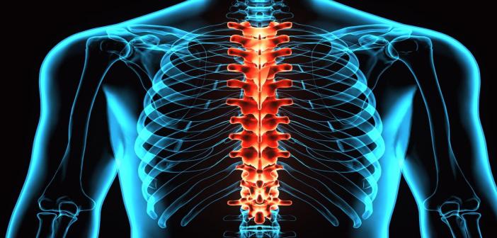
The cervical, thoracic, and lumbar spines are commonly divided into three main parts to illustrate the spine's normal anatomy. (The sacrum is a member of the pelvic girdle, is just behind the lumbar spine.) Individual bones are called vertebrae, make up each portion. Seven cervical vertebrae, twelve thoracic vertebrae, and five lumbar vertebrae make up the spine.
Brief about the spinal anatomy
Multiple sections make up a single vertebra. The main weight-bearing field for the vertebrae in the body also serves as a resting place for the fibrous discs that distinguish them. The vertebral canal, a wide opening in the middle of the vertebra into which the spinal nerves travel, is covered by the lamina.
When you run your hands down your back, you can feel the spinous process which gives you an idea about the spinal anatomy. Back muscles are attached to the paired transverse processes aligned 90 degrees to the spinous process. Each vertebra is connected with four facet joints. There are two pairs, one facing upward and the other facing downward.
The interlocking of these vertebrae with the neighboring ones gives the spine cohesion. Intervertebral discs serve as cushions in the middle of the bones and differentiate the vertebrae. Each disc has two parts. The annulus is a rigid, rugged outer layer covering the nucleus, which is mushy and sticky.
The elastic nucleus of a herniated or ruptured disc spurts out via a rupture in the annulus, compressing a nerve root. The nucleus will squirt out of the disc on either side or both sides in some situations.
The number of nuclei that penetrate the annulus and where it suffocates a nerve. Determines how much damage a disc rupture causes. A laminectomy/microdiscectomy procedure can be used to help relieve the discomfort.
Stabilizers and Enablers for Spine Movement
The spinal column is made up of more than just bones. The spine depends on various supportive mechanisms to retain its double-S form, provide skeletal support, and route nerves where they need to go.
The spinal discs, a part of spinal anatomy, also known as intervertebral discs, are the first of these systems. From the C3 to the L5-sacrum, each disc resembles a fibrous tissue pad (called fibrocartilage) and is held in place with the help of vertebral endplates (called cartilaginous endplates). These discs serve as shock absorbers and interbody spacers. Neither there is a disc between C1 and C2 nor between sacrum and coccyx.
Behind the vertebral body, facet joints are combined (left and right sides) (C3-L5). These joints are also known as zygapophyseal or apophyseal joints.
These joints aid in the stabilization of the spine while enabling flexion (bending forward), extension (bending backward), and twisting movement (called articulation).
Each facet joint is encased in a connective tissue capsule that contains a nourishing fluid to lubricate the joints, similar to other joints in the body. The smooth mobility of the joints is ensured by cartilage coating the joint surfaces.
Spinal Nerves protected by the Vertebral Column
The central nervous system includes the spinal cord and nerve roots. This begins at the bottom of the skull, which is protected by the vertebral column. From the cervical to the lumbar spine, the vertebral systems form a single round hollow area that contains the spinal cord.
A foramen, also known as foramina, is where small nerve roots arise from the spinal cord and exit the vertebral column. An intervertebral disc anchored between two vertebral bodies creates each foraminal space on the left and right sides. Since exiting the spinal column, the nerve root branches into the peripheral nervous system, which serves the whole body from head to toe.


0 Comments